User Information
Request SIMS time
Maximizing your productivity
Hourly Fees
Notice
Request SIMS time
Time on the ASU SIMS instruments can be requested by email. Please send your request to sims@asu.edu. Be sure to include:
- Your name and affiliation.
- A short description of the project which MUST
include:
- the elements or isotope ratios you wish to determine
- the lateral resolution required for each analysis
- the precision required for each analysis
- the approximate number of analyses you hope to obtain.
- Optimum dates for your session.
- How many people would be in your visiting group.
In addition, please tell us if NSF or another funding agency funds the project.
Information supplied by these questions can help the SIMS lab personnel prepare
to help you get the most out of your analysis session (also see below).
Maximizing your productivity
The best visits to any SIMS lab occur when the visitor considers carefully
these questions:
How should I prepare/mount my samples?
How much time do I need for each analysis?
The two important parts of sample preparation include sample mounting (especially for analyzing "volatile" elements) and photodocumentation so that you can easily locate areas for analysis. These topics are described below.
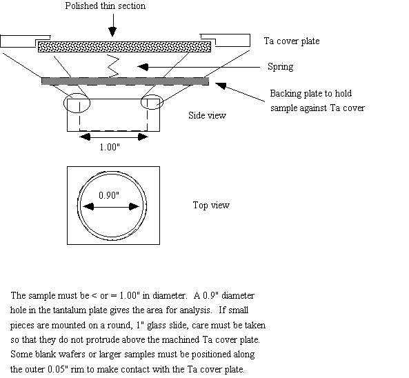
Sample Mounting. The Cameca SIMS instruments we work with can hold one 1" diameter sample with a maximum thickness of ~1/2" (see figure below). Sample changes require a minimum of about 5 minutes, so it is feasible to analyze several samples during a session. However, many outside users wish to analyze for H in various phases and hydrogen background signals may take hours to decay to baseline conditions after a sample change. In addition, some mounting media (various epoxies) degas to a large extent, requiring a much longer time to change samples (read more about epoxy below). Thus, it is beneficial to mount as many samples as possible on each 1" diameter plug. Please note that (except in special circumstances) not all of the sample surface can be analyzed. The edge of the sample is not accessible for study. How close to the edge one can get depends on a number of factors, but we suggest eliminating the last 0.1" (or more) from consideration. Requests to analyze phases at the edge of a polished, round, 1" diameter thin section must be made separately.
Minimizing the hydrogen background.
Multiple samples can be mounted in a prepared disk of epoxy, or onto a
1" diameter
round glass slide (with minimal epoxy). All epoxies will degas somewhat,
and this local source of hydrogen seems to represent the most important
contribution to the H background signal. In the 3f SIMS, we have observed
the lowest H background (corresponding to ~0.1 wt.% H2O) when we analyze
thin wafers (suitable for infrared spectroscopic analysis) that are glued
to a glass slide with a drop of "superglue" or pressed
into double-sided (vacuum compatible) sticky tape.
For the lowest levels of detection, users press polished samples into indium metal. The polished, flat sample allows reproducible analyses to be obtained while the indium metal does not contribute a hydrogen signal to the vacuum system. This mounting style is suitable for the analysis of hydrogen in "nominally anhydrous minerals" (NAMs) and other samples with low water contents (e.g., apatites, volcanic glasses quenched at low pressures). It is also useful for applying depth profiling techniques to mineral surfaces (for example, measuring diffusion profiles where the diffusing species has penetrated less than a few hundred to few thousand nanometers into the sample surface)
Sample Polishing. SIMS is a surface analytical technique, so sample preparation is extremely important. Anything that is left on the sample surface may be analyzed during your session. This includes residual polishing media, the conducting coat deposited on the sample, and inadvertent contamination. We recommend that visitors consider the elements they want to study before final sample preparation. For example, applying a carbon coat would be clearly inappropriate for measuring carbon, and polishing with fine-grained alumina may cause problems in a subsequent analysis for Al in quartz (or oxygen isotope microanalysis). One of the most serious contamination problems observed is for boron. Boron contents of polishing materials may be high, or it may settle on a sample as dust (this can be very important). We have also observed that grains that are placed in vacuum grease (to hold them while epoxy is poured over them) can have enormously increased signals for Li. We strongly recommend that when making grain mounts, the isolated grains be placed on double-sided tape prior to adding epoxy to minimize such contamination.
Locating areas for analysis. We cannot emphasize enough the poor quality of the optical microscope attached to the SIMS (although the 6f microscope is better than the 3f). Visitors are required to bring low-magnification (≤50X preferred), reflected light images of the areas of interest. Images obtained on a scanning electron microscope are sometimes useful navigation aids, and are best if taken in secondary electron imaging (SEI) mode at low resolution. If the automatic scale bar in such an image is below 100 microns, then the image is probably too high in resolution to be useful in locating areas of interest. We repeat: low-magnification reflected light images are the best. Transmitted light and cross-polarized transmitted light images are petrographically useful, but may not help you find areas for analysis. If you bring inappropriate images to the lab we will send you and your samples to an optical microscope equipped with a digital camera where you will obtain useful images. Please note that we will charge you for this time.
Hourly Fees
The ASU SIMS labs have no protocol for performing proprietary work; there are several consulting firms eager to perform such analyses. We currently charge an hourly fee (minimum charge of 4 hours) for SIMS time: $35/hour on the 3f and $60/hour on the 6f SIMS. Please see our Applications page for particular expertise of the ASU lab.
Notice
Arizona State University is not responsible for any injury to non-ASU personnel that occurs on ASU property. Visitors to the lab are covered by their existing insurance only.